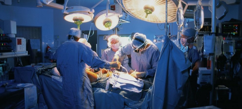
LSG mechanisms LSG involved in the GLP-1 secretion mechanism

Laparoscopic sleeve gastrectomy accelerates intestinal motility and reduces intestinal transit time, which may be involved in the mechanism underlying enhanced glucagon-like peptide-1 (GLP-1) secretion during oral glucose tolerance test (OGTT) after the procedure, according to paper by researchers from Shiga University of Medical Science, Otsu, Shiga, Japan.
The study, ‘Enhanced Intestinal Motility during Oral Glucose Tolerance Test after Laparoscopic Sleeve Gastrectomy: Preliminary Results Using Cine Magnetic Resonance Imaging’, published online in the journal Plos One, showed that GLP-1 secretion during OGTT was enhanced and cine magnetic resonance imaging (MRI) showed markedly increased intestinal motility at 15 and 30 min during OGTT after laparoscopic sleeve gastrectomy.
Although enhanced secretion of GLP-1 has been suggested as a possible mechanism underlying the improvement in type 2 diabetes mellitus (T2DM) after sleeve gastrectomy, the reason for enhanced GLP-1 secretion during glucose challenge after the procedure remains unclear because the procedure does not include intestinal bypass.
Study
Therefore, the study authors focused on the effects of the procedure on GLP-1 secretion and intestinal motility during the OGTT using cine MRI before and three months after LSG. They used this specific imaging modality as it “provides direct visualisation of intestinal contraction and peristalsis”.
Twelve obese patients with a BMI>35 were recruited into the study; six patients had T2DM and two diabetic patients had haemoglobin A1c (HbA1c) levels >7.8%.
Sleeve gastrectomy was performed using a standard five-port laparoscopic technique with a 45-Fr gastric tube to calibrate the sleeve, and dissection of the greater curvature began approximately 5–6 cm from the pylorus.
MRI examinations were performed for each patient one week before and three months after the surgery and MRI was conducted after eight hours of fasting before as well as 15 and 30 minutes after oral intake of 225mL of fluid containing 7 g of glucose.
Imaging was performed using a 1.5T MR scanner (Signa HDxt 1.5T; GE Healthcare,) with an 8-channel body array coil. Before real cine MRI, coronal images of the entire abdomen were obtained to determine the optimal image plane covering the maximum length of the small bowel loops. A serial coronal scan consisting of 50 images was obtained at the selected plane with the patient in a supine position in 25 seconds during breath holding.
Based on the cine MRI, 2 bowel segments, one located in the left upper quadrant as representative of the jejunal loops and the other located in the right lower quadrant as representative of the ileal loops, were chosen for assessment of contraction
In this process, bowel loops with a degree of distension similar to the rest of the loops in the same quadrant as well as remaining in the image plane during the sequential imaging without displacement out of the image plane were chosen for assessment. Frequencies of bowel contractions were counted visually on a monitor using cine MRI.

